Brain - Movement
MOVEMENT
This work was done as a part of Summer Program at Elio Academy of Biomedical Sciences.
Author: Riyaa Sri Ramanathan
The central nervous system — brain and spinal cord — directs the coordinated actions of the hundreds of muscles that enable humans to move.
The nervous system governs motion by controlling the structures of the body that produce movement — the muscles. Most muscles attach to the skeleton and span joints, the sites where two or more bones come together. The relationship of the muscles with the skeleton is called skeletal muscles. Activating muscles can either flex or extend the joint that they span. Muscles that bend a joint, bringing the bones closer together, are called flexors; muscles that straighten the joint, increasing the angle between the bones, are called extensors.
Flexors and extensors work in opposition, so when one set of muscles contracts, the other relaxes. For example, bending the elbow requires contraction of the biceps (a flexor) and relaxation of the triceps (an extensor). For such motions, the muscles that promote the movement are called agonists, and those that oppose or inhibit the movement are antagonists. Skilled, rapid movements — like throwing a dart — are started by agonists and stopped by antagonists, allowing the limb to accelerate and halt with great speed and precision. For some movements, agonists and antagonists contract at the same time, which is called co-contraction.
These simultaneous actions can stabilize or control a movement in different cases or scenarios. Whether flexion or extension, the movement of all skeletal muscles is controlled by the central nervous system. A skeletal muscle is made up of thousands of individual muscle cells, called muscle fibers. Each muscle fiber is controlled by a single alpha motor neuron that originates in the spinal cord or the brain. However, each of these alpha motor neurons can control a few to hundred or more multiple muscle fibers.
An alpha motor neuron + all of the muscle fibers it controls forms a functional unit known as the motor unit, the critical link between the central nervous system and skeletal muscles. When motor neurons die, in diseases like amyotrophic lateral sclerosis (ALS), people can lose their ability to move as well. Some muscles act on soft tissues, but not on joints. For example, muscles in the head and neck enable the eyes to move, chew and swallow food, have conversations and control facial expressions. These muscles are also controlled by the central nervous system (CNS), and operate the same way as those attached to bones.
Involuntary movements mostly take place without conscious control. The most fundamental types of voluntary movements are the reflexes, which are stereotyped, automatic muscle responses to particular stimuli. These reflexes get involved in the activation of sensory receptors in the skin, joints, or muscles. The responses are rapid and occur without the involvement of the brain or conscious attention. Instead, they depend on circuits of neurons located in or near the spinal cord.
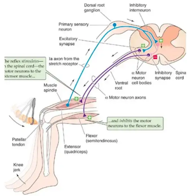
The best known reflexes is the “knee jerk response”,a stretch (myotatic) reflex that occurs when a physician strikes the tendon just below the knee with a small rubber hammer. This tap produces slight stretch of the knee extensor muscle, which is “sensed” by receptors within the muscle called muscle spindles. The spindles sense the extent and speed of the stretch, and stimulate sensory neurons, which send impulses into the spinal cord. There, the signals activate the alpha motor neurons that cause the stretched extensor muscle to contract, triggering the reflex. However, for the leg to kick forward, the antagonist flexor muscle has to relax at the same time. The same sensory stimuli that directly activates the motor neurons controlling the extensor also indirectly inhibits the motor neurons controlling the antagonist flexor. This reciprocal inhibition is accomplished by connecting neurons that lie completely within the spinal cord.
When these inhibitory interneurons are activated by the original sensory stimulus, they send impulses that inhibit the motor neurons supplying the flexor. Thus, even the simplest of reflexes involves the synchronous activation, and inactivation of multiple sets of motor neurons controlling both agonist and antagonist muscles. Many reflexes protect from injury. One such reflex is the flexion withdrawal reflex that occurs when barefoot encounters a sharp object. In this case, pain receptors in the skin send a message to the spinal cord, alpha motor neurons are activated, and the leg is immediately lifted. At the same time, as the body weight is supported on both legs, the extensors of the opposite leg must be activated. Without this additional reaction, called the flexion crossed extension reflex, loss of balance would occur. When these movements occur, the muscles involved provide feedback to the brain with information about where the various body parts are in space and how fast they are moving. The muscle spindles mentioned above supply information about changes in muscle length or stretch. In turn, the brain adjusts the sensitivity of the system via a separate set of motor neurons and gamma motor neurons which keeps the muscles tensed. Other specialized receptors called Golgi tendon organs, located where the muscle fibers connect to the tendon, detects how much force or tension is applied to a muscle during ongoing movement by increasing the movement’s precision. The golgi tendon organ receptors are located where the muscle fibers connect to the tendon.
Voluntary behaviors are mainly controlled by spinal circuits. Locomotion which occurs during the rhythmic patterns of muscle activation are generated when neurons get activated within the spinal cord and brainstem circuits. The movements that are triggered include walking, flying, swimming, or breathing etc. Central pattern generators which evolved in primitive vertebrates are being studied to determine the level to which spinal circuitry can be co-opted to recover basic postural and locomotor function after severe paralysis. The most complex movements that the body performs, including those requiring conscious planning, involve input from the brain. These higher brain regions initiate voluntary motion, coordinate complex sequences of movement, and support behavioral output for a given situation. These programs expect the brain to relay commands to appropriate spinal circuits. Scientists are just beginning to understand the coordinated series of interactions that take place among different brain regions during voluntary movement. One brain area essential for voluntary movement is the motor cortex. Neurons in the motor cortex send signals that directly control the activation of alpha motor neurons in the spine. Some of these cortical neurons control the movement of related muscles in an individual body part such as a hand or arm, while other neurons in the motor cortex direct the coordinated movement of a limb to a particular position.
Complex body movements are controlled by different brain regions which include the motor cortex, several brain regions that participate in parallel circuits or loops, the basal ganglia, thalamus, cerebellum and a large number of neuron groups located within the midbrain and brainstem. These regions also influence the activity of motor neurons in the spine. The basal ganglia itself surrounds two separate pathways — one to facilitate the desired motor program while the other to suppress the unwanted, competing actions. Along with the thalamus, the basal ganglia share connections with motor and sensory areas of the cerebral cortex, allowing these structures to monitor and adjust motor performance.
Dysfunction of basal ganglia can lead to severe movement disorders. People with Parkinson’s disease experience degeneration of neurons in a brain region called the substantia nigra. These neurons relay signals to the basal ganglia using the neurotransmitter dopamine, a key chemical involved in motor control. Depletion of dopamine results in the symptoms of PD including tremor, rigidity, and in some cases, akinesia, an inability to move.
Individuals with Huntington’s disease often display uncontrolled jerking or twitching movements, particularly in the face and extremities. These symptoms result from a selective loss of inhibitory neurons in the basal ganglia, which eliminates the suppression of random involuntary movements. Another brain region crucial for coordinating and fine-tuning skilled movement is the cerebellum. The cerebellum receives direct input from sensory receptors in the limbs and head, as well as most areas of the cerebral cortex.
Neurons in the cerebellum integrate the sensory information to ensure proper timing and integration of muscle action. This enables the body to produce fluid movements more or less automatically. The cerebellum is essential for a wide range of motor learning and coordination, from controlling limb movements to eye movement to grip force. Disturbance of cerebellar function leads to poor coordination, disorders of balance, and even difficulties in speech, one of the most intricate forms of movement control. Long Term alcohol abuse is a common cause of acquired cerebellar degeneration.
Typical symptoms are poor coordination, an unsteady walk or stumbling gait, changes in speech, and difficulty with fine motor skills including eating, writing etc. The Cerebellum also allows humans to adapt to unexpected movements, helps to recalibrate motion as body’s changes in height, weight, disease or disability, and also facilitates skillful movements as humans grow by age.
Author: Riyaa Sri Ramanathan
This work was done as a part of Extended Research Program at Elio Academy of Biomedical Sciences.


Comments
Post a Comment